
X-ray before treatment: teeth with caries and unexact crowns, chronical periapical inflammation on both roots
X-ray after removal of crowns, build-up and root canal retreatment
Control X-ray – new crowns, healed periapical inflammation
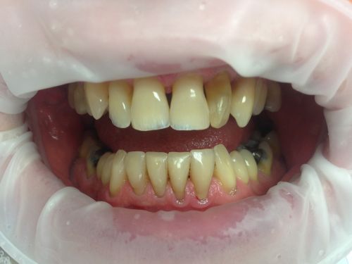
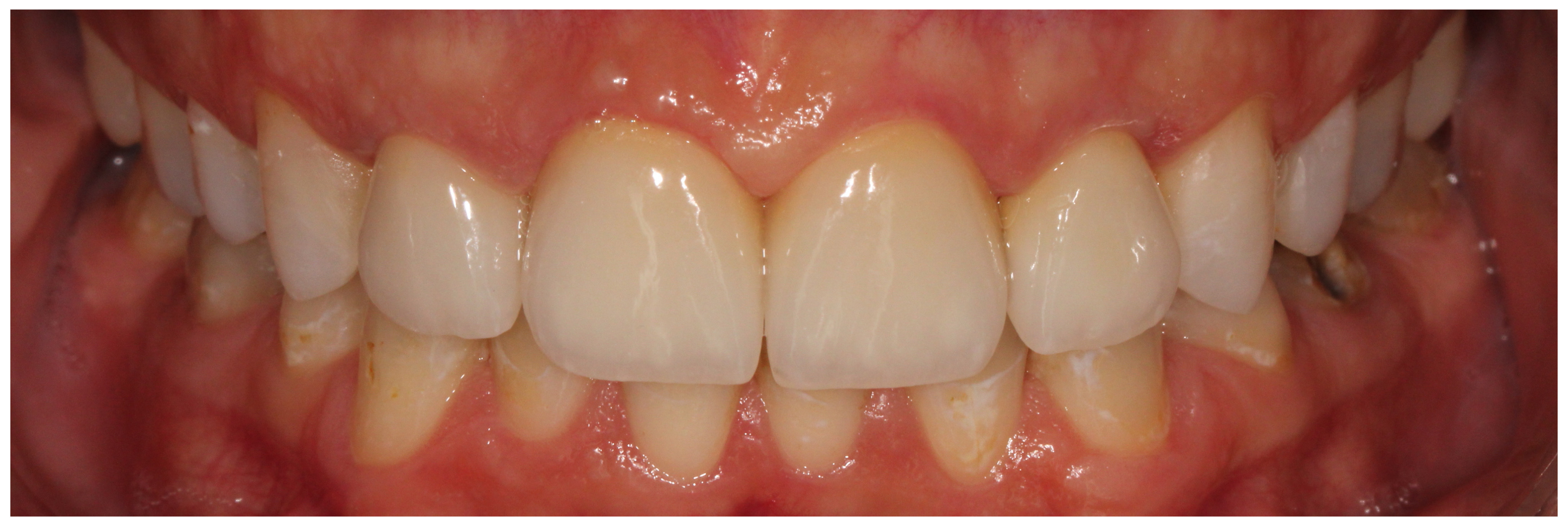
Before teeth whitening and smile correction on patient's request
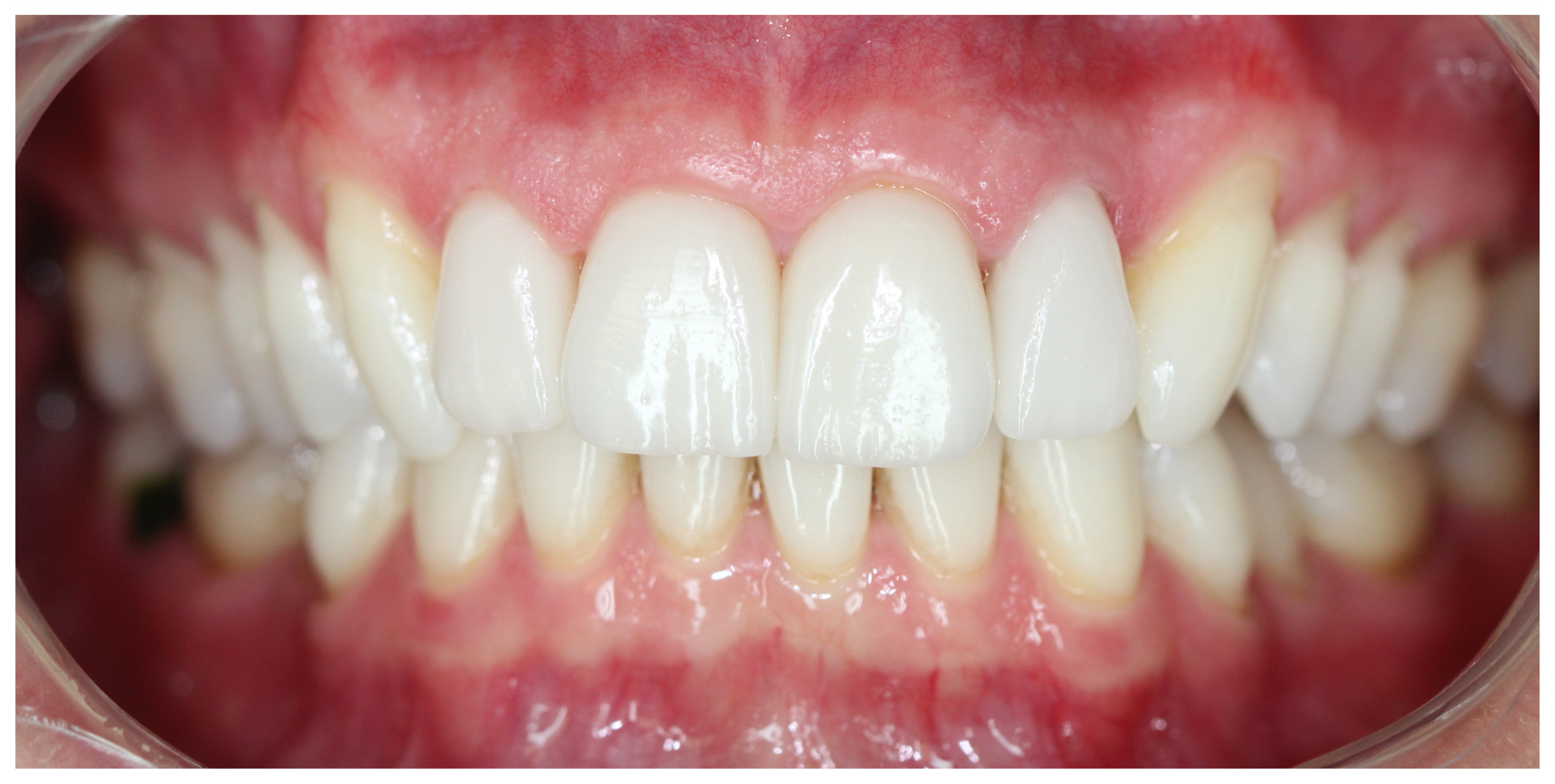
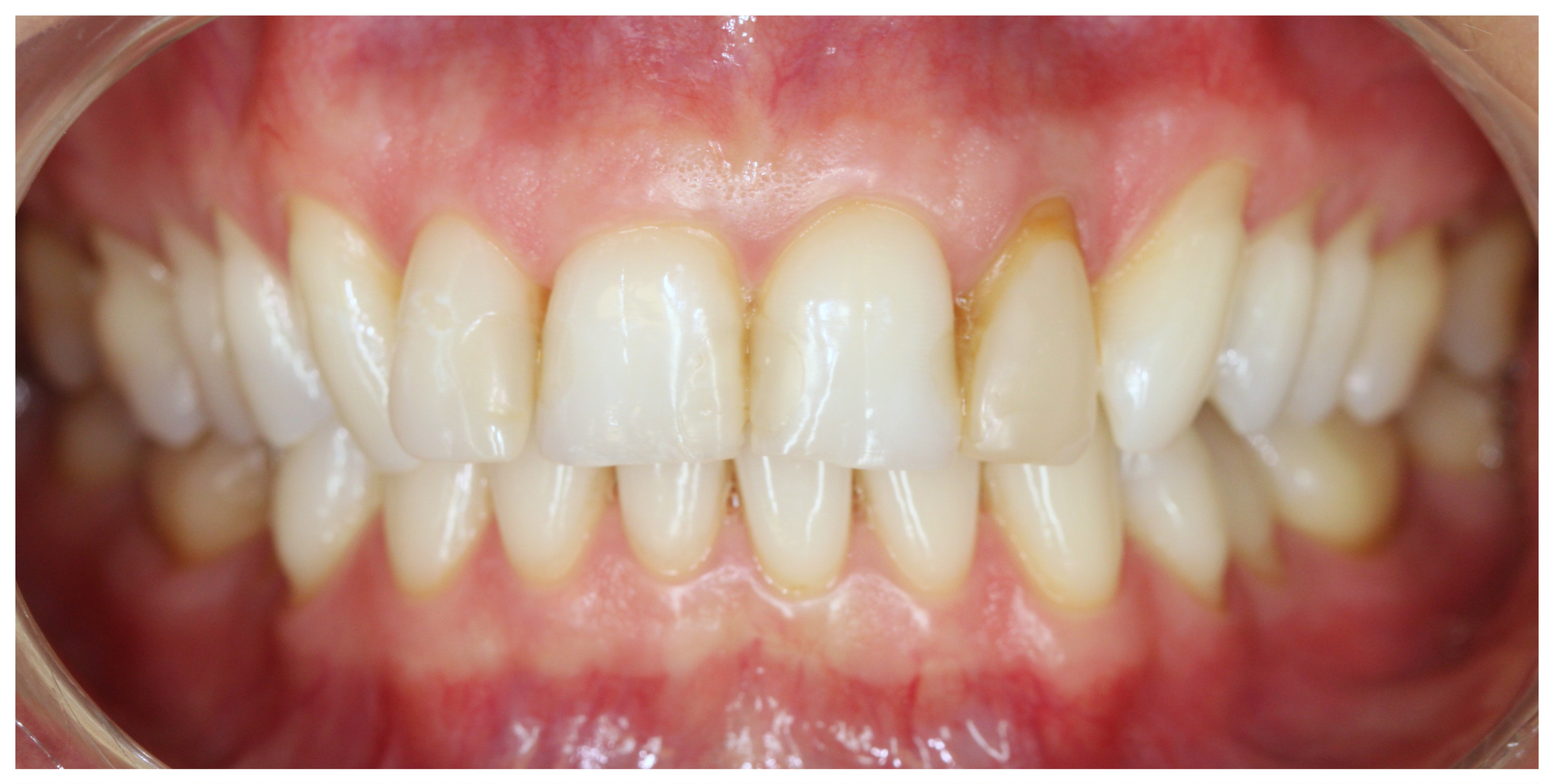
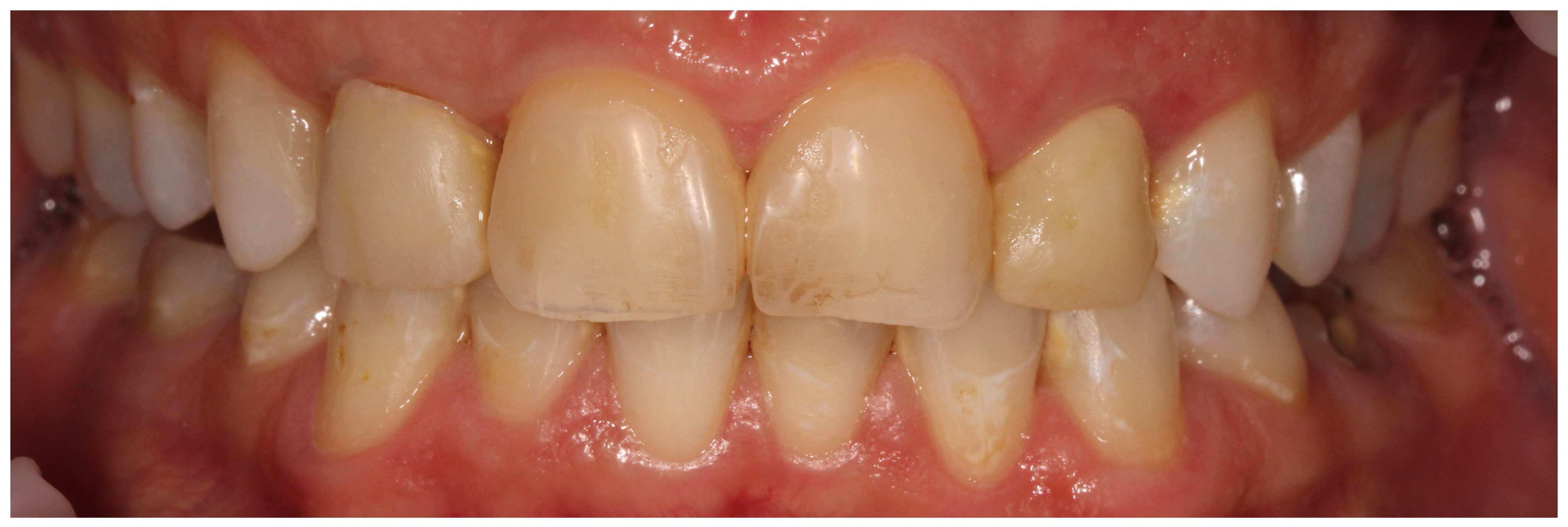
After teeth whitening and with three new ceramic veneers and one crown on upper incisors
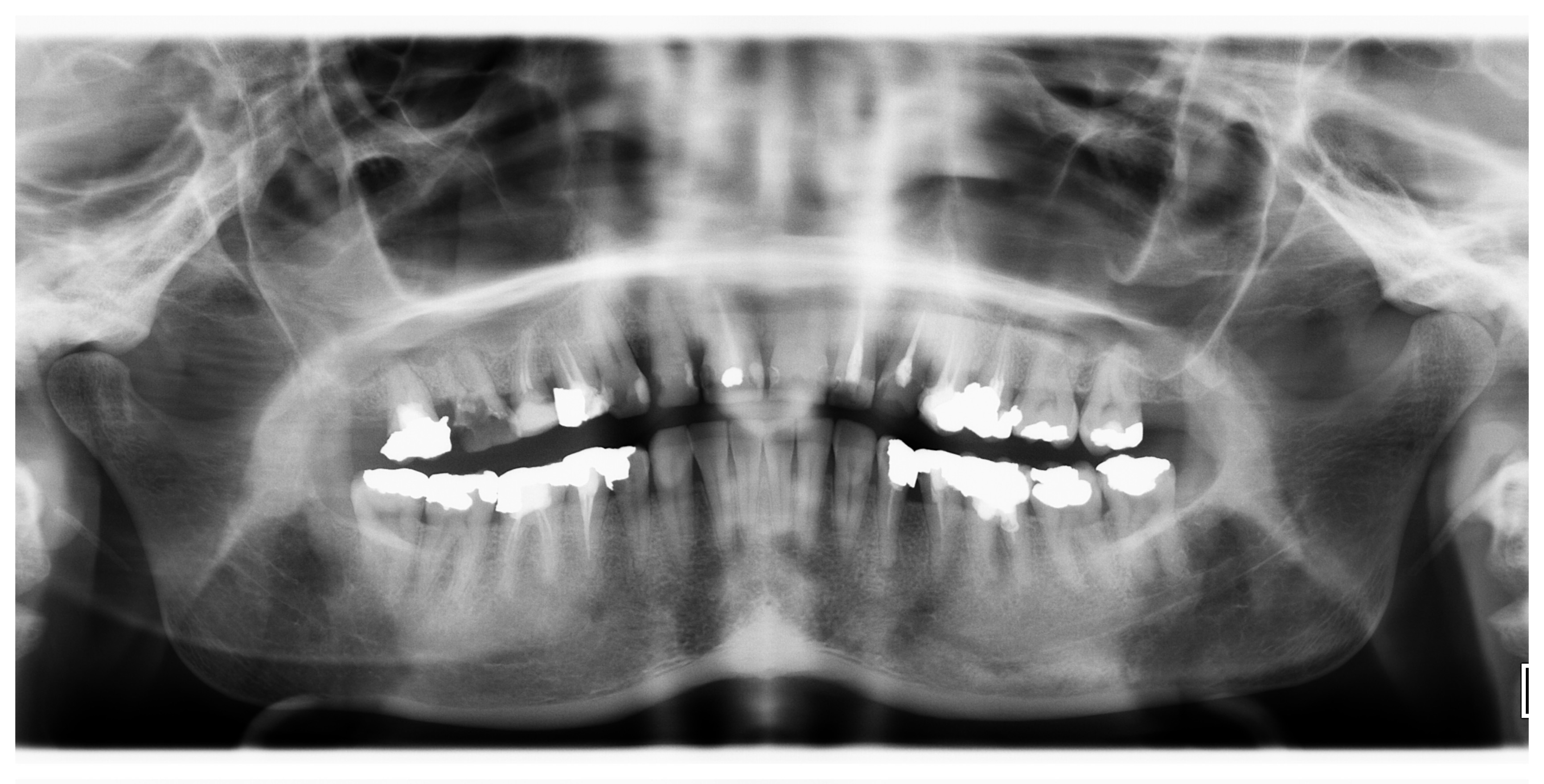
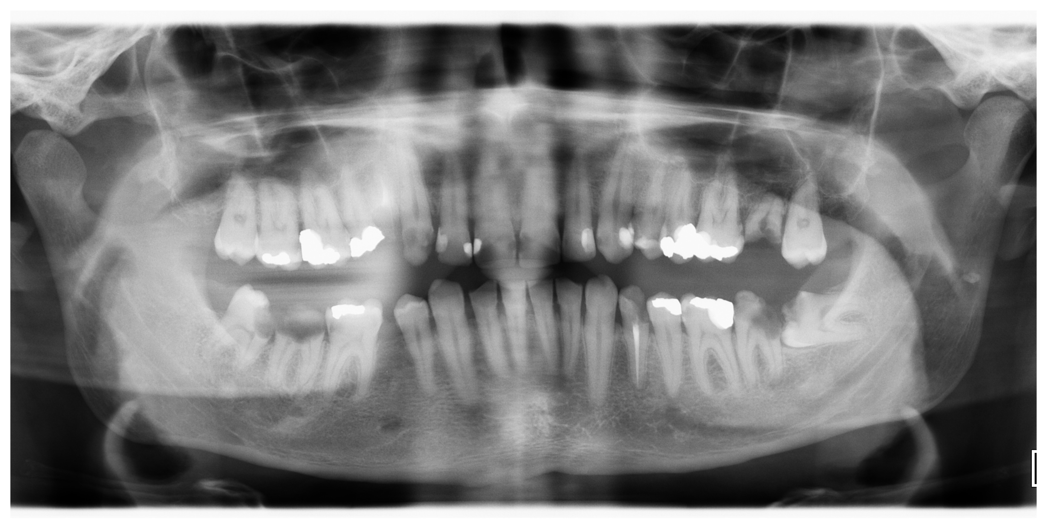
Panoramic X-ray during initial consultation – multiple caries under unexact fillings, periapical inflammation, inadequate root canal treatments
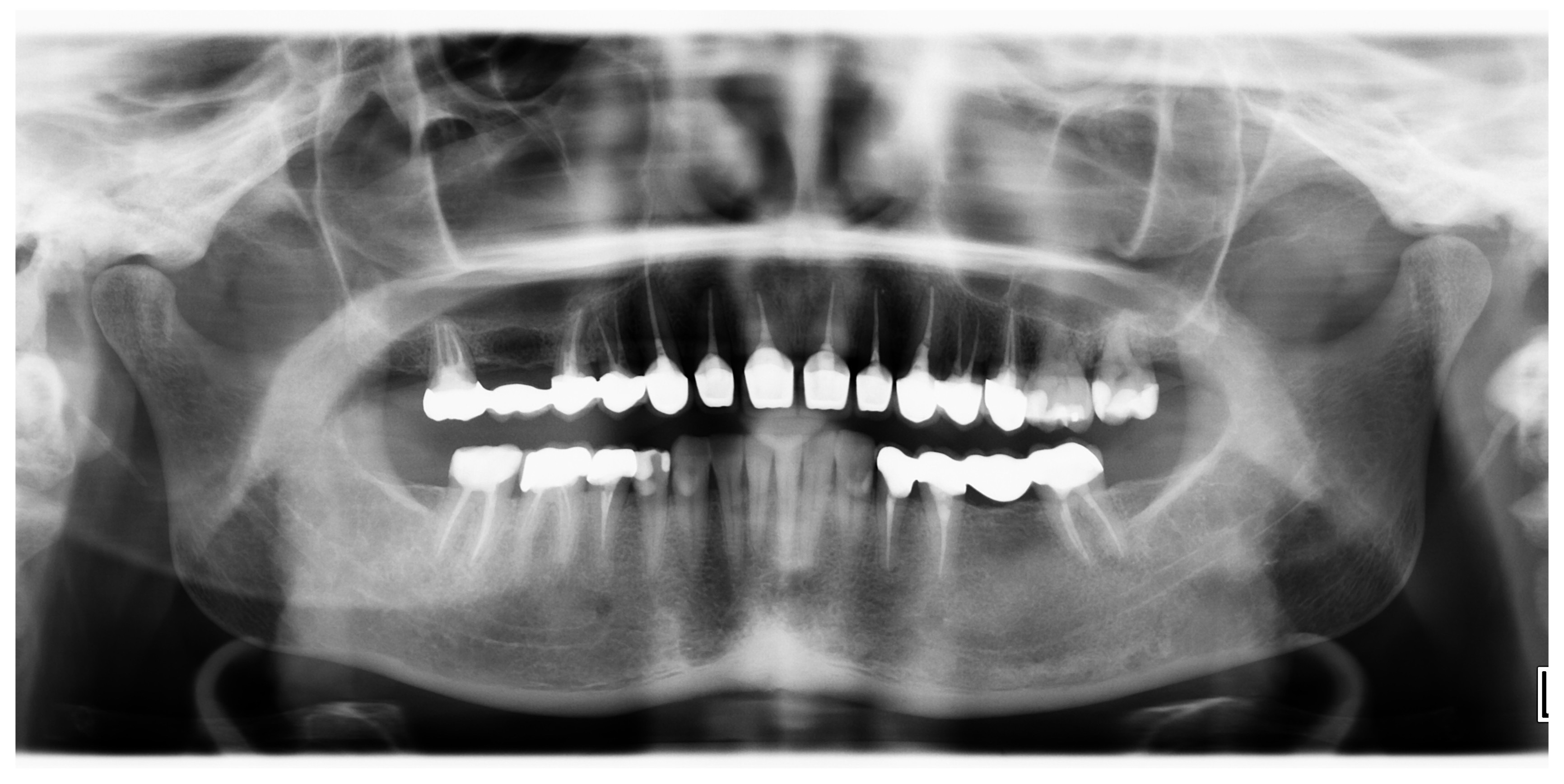
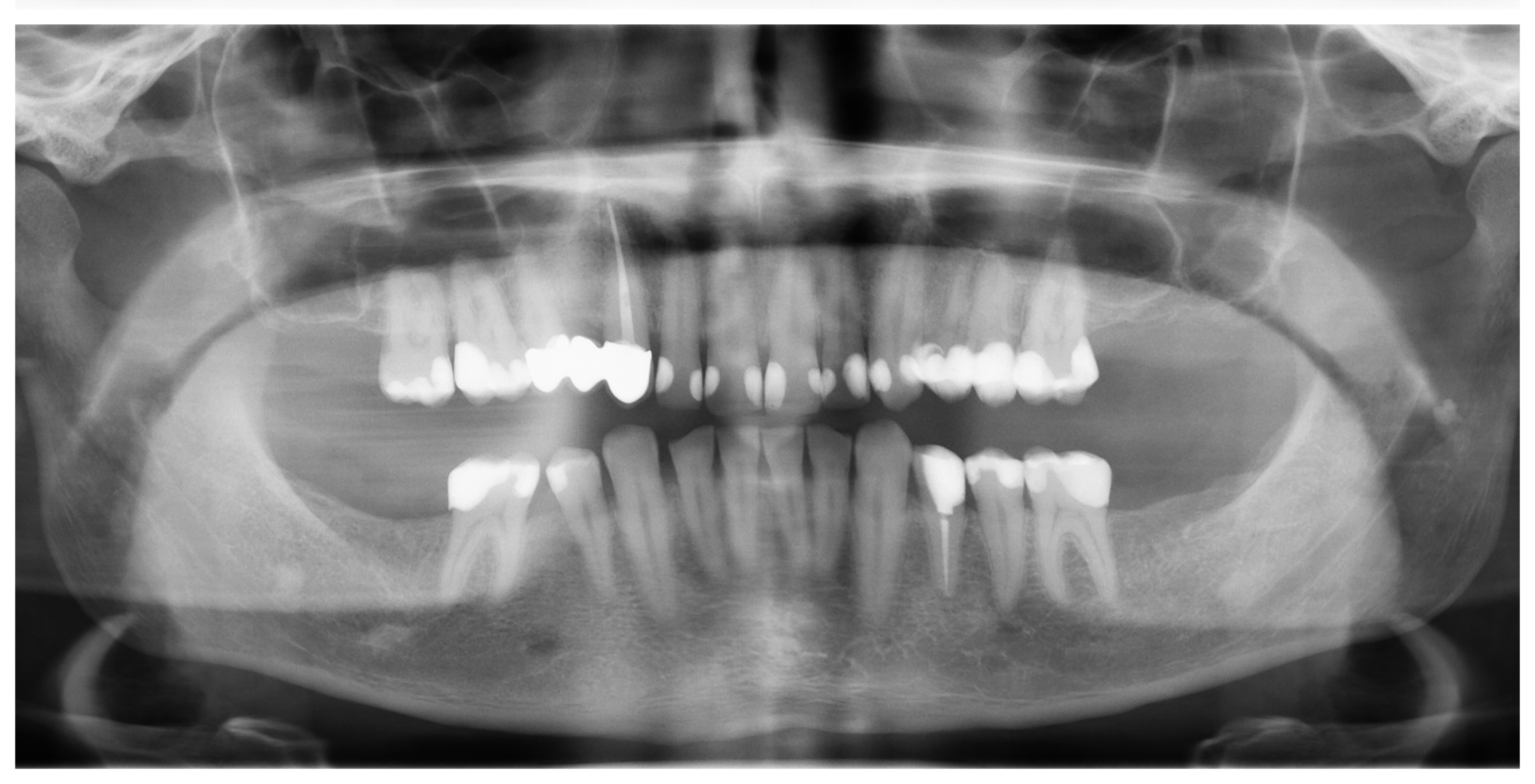
Panoramic X-ray made three years after termination of treatment – new root canal treatments, new precise crowns and bridges, patient without any pain or other problems
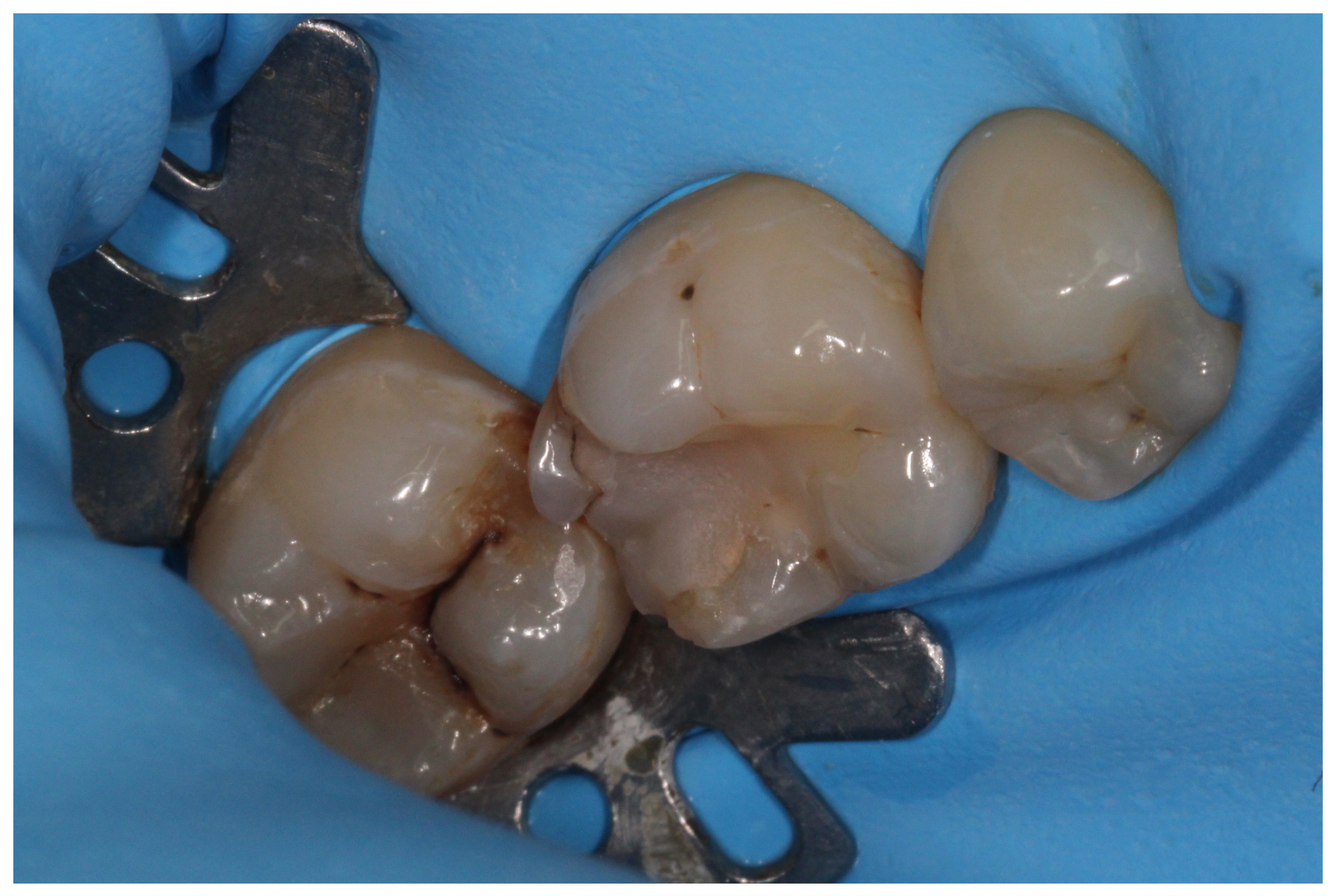
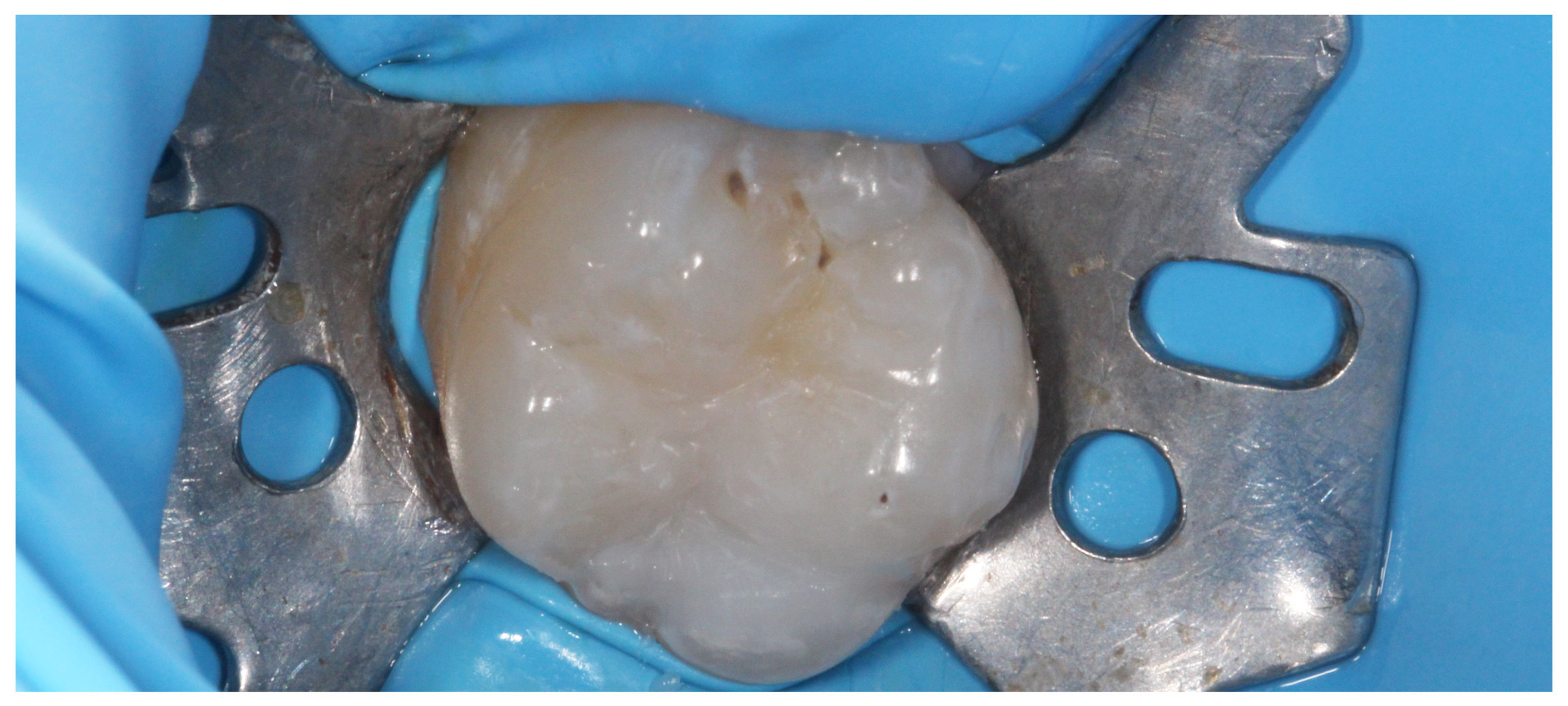
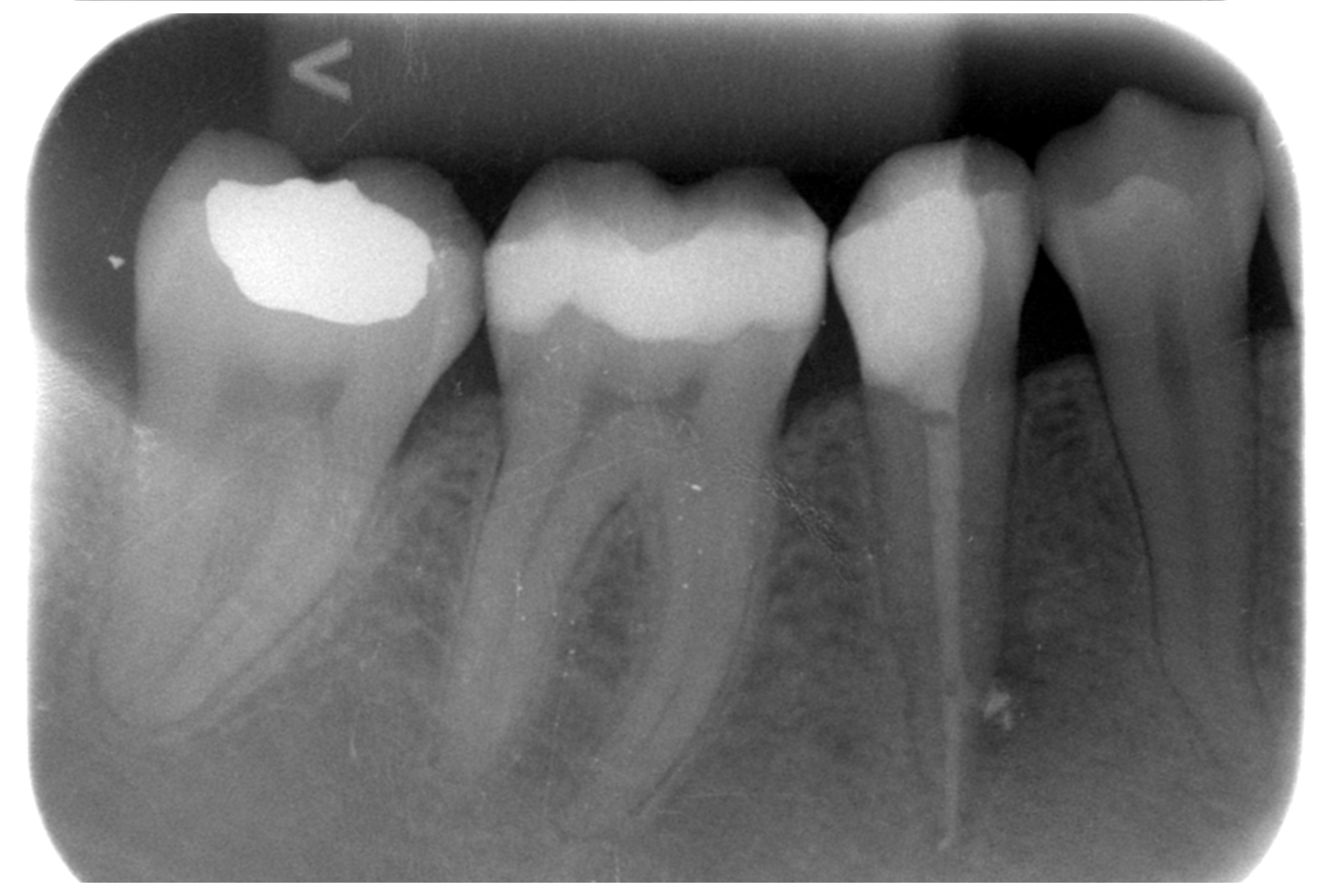
„White“ filling with caries on the first molar, new caries on the second molar
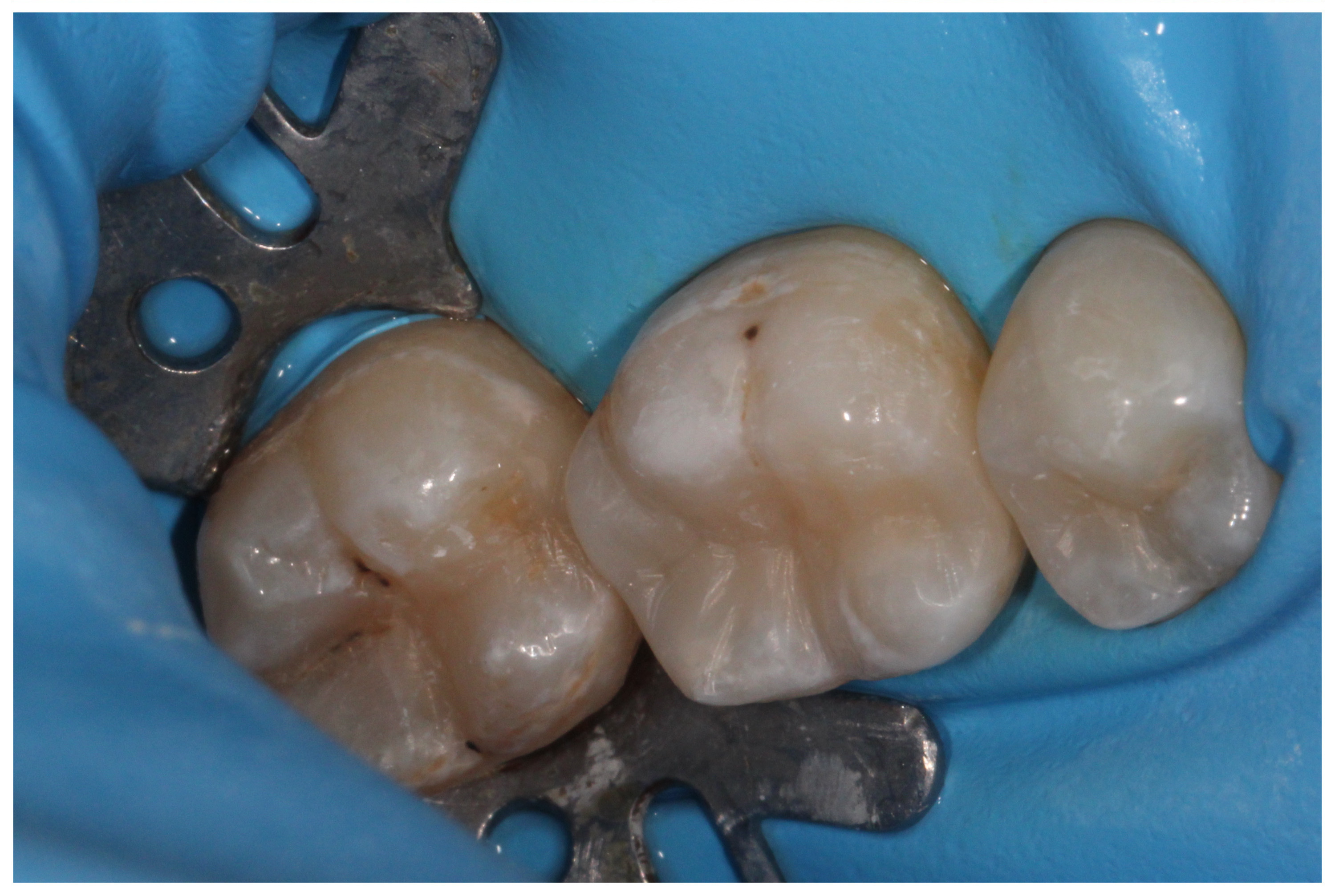
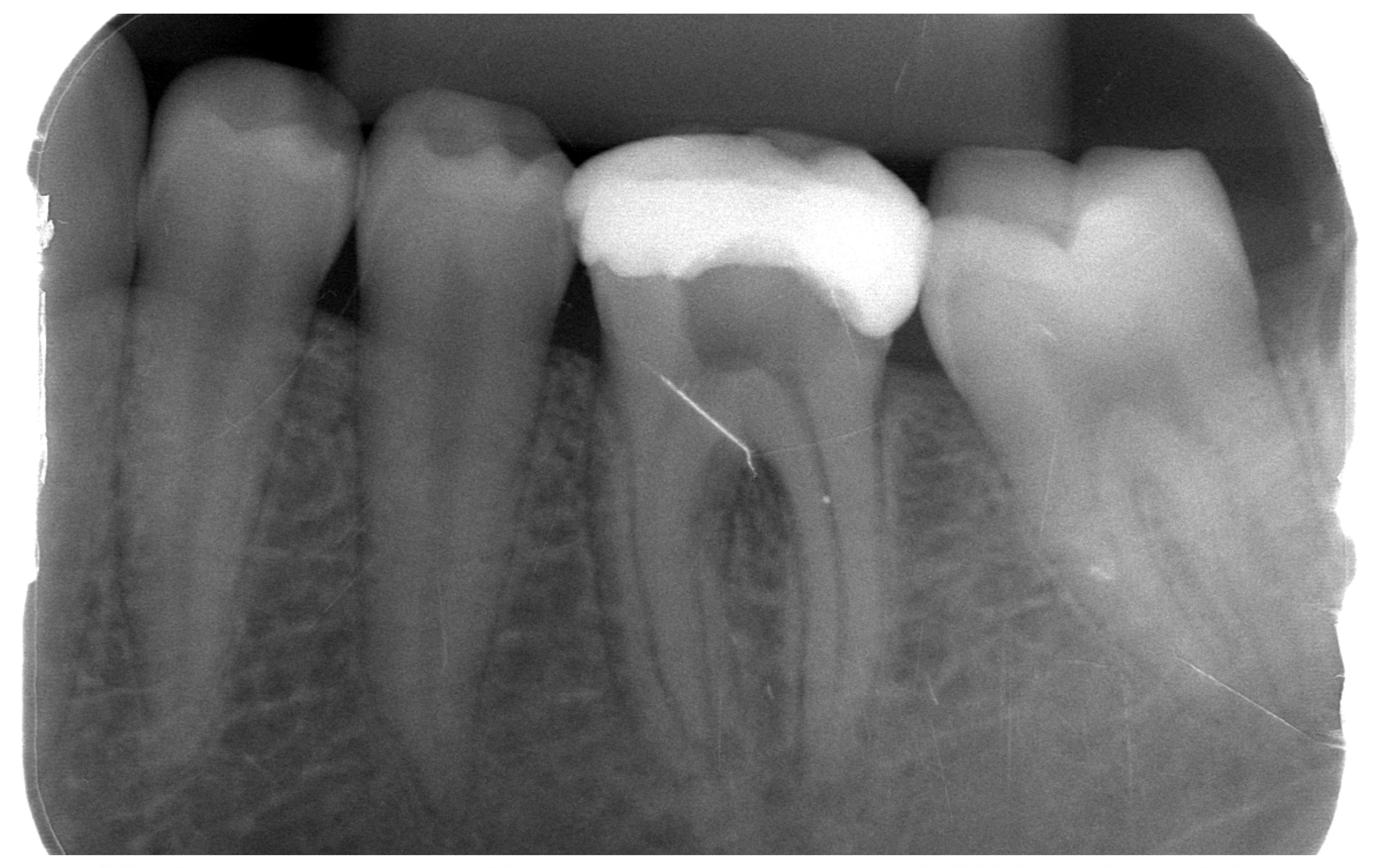
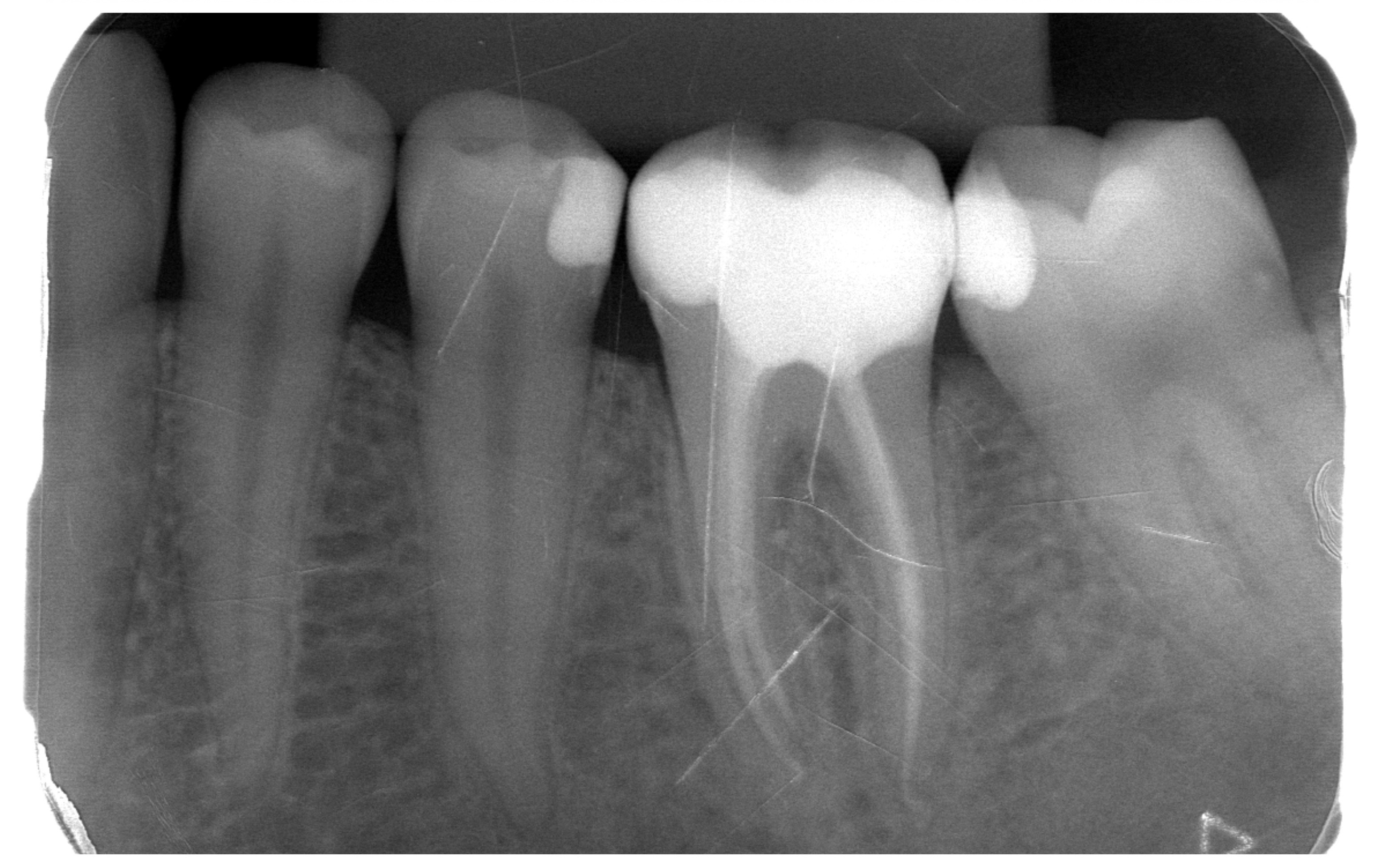
New photocomposite fillings
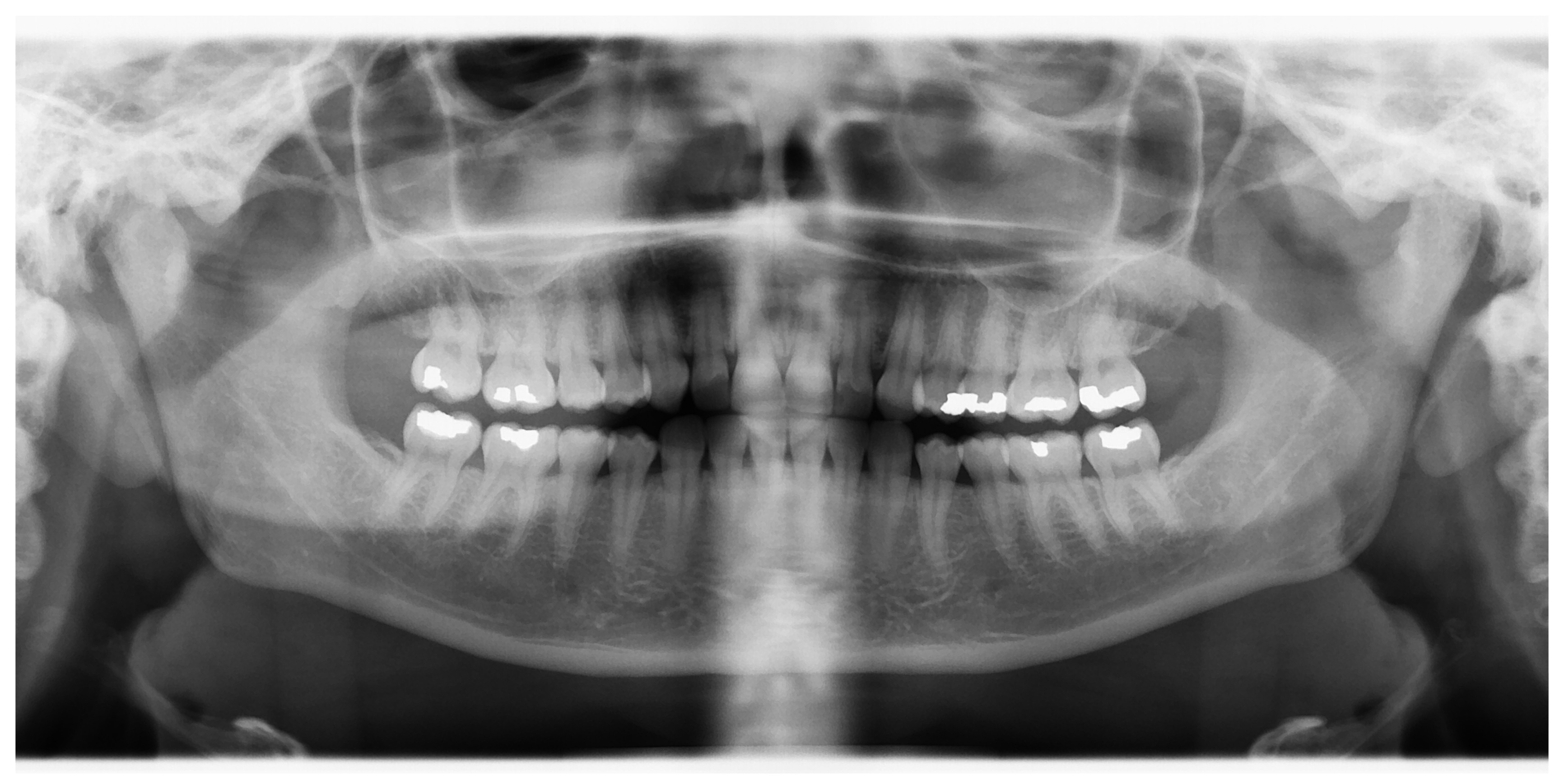
Panoramic X-ray during initial consultation – new caries and caries under old fillings
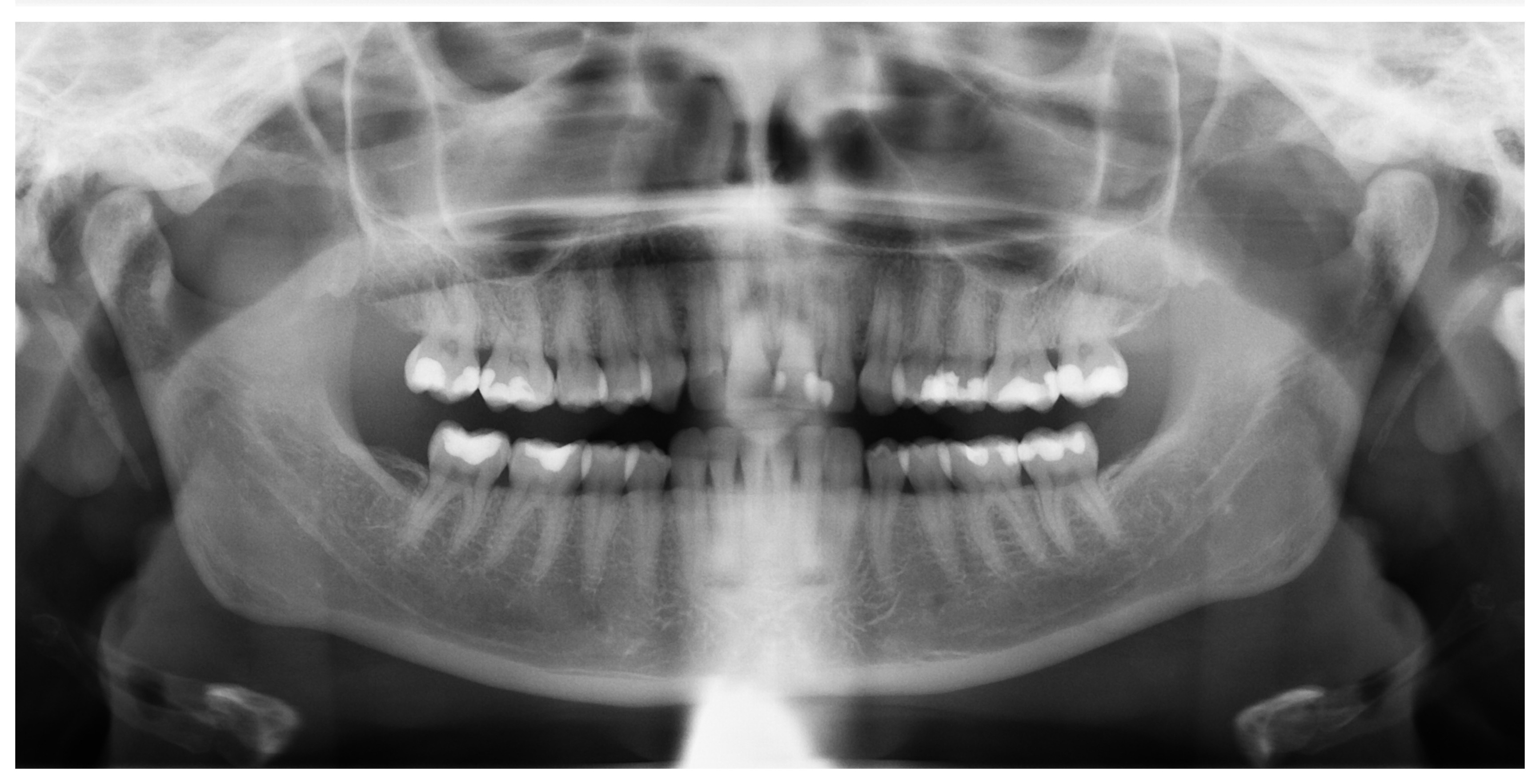
Control panoramic X-ray two years after termination of treatment
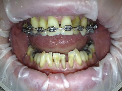
Excessively worn teeth because of orthodontic anomaly
Direct composite build-ups after orthodontic treatment
Panoramic X-ray during initial consultation – multiple caries and remaining roots for extraction
Control panoramic X-ray three years after termination of treatment – long-term stable result, bone after mutliple extractions fully healed, caries treated, space after extraction of upper premolar solved with bridge
Provisional crowns on upper second incisors, large photocomposite build-ups on first incisors
Permanent full-ceramic crowns on upper first and second incisors
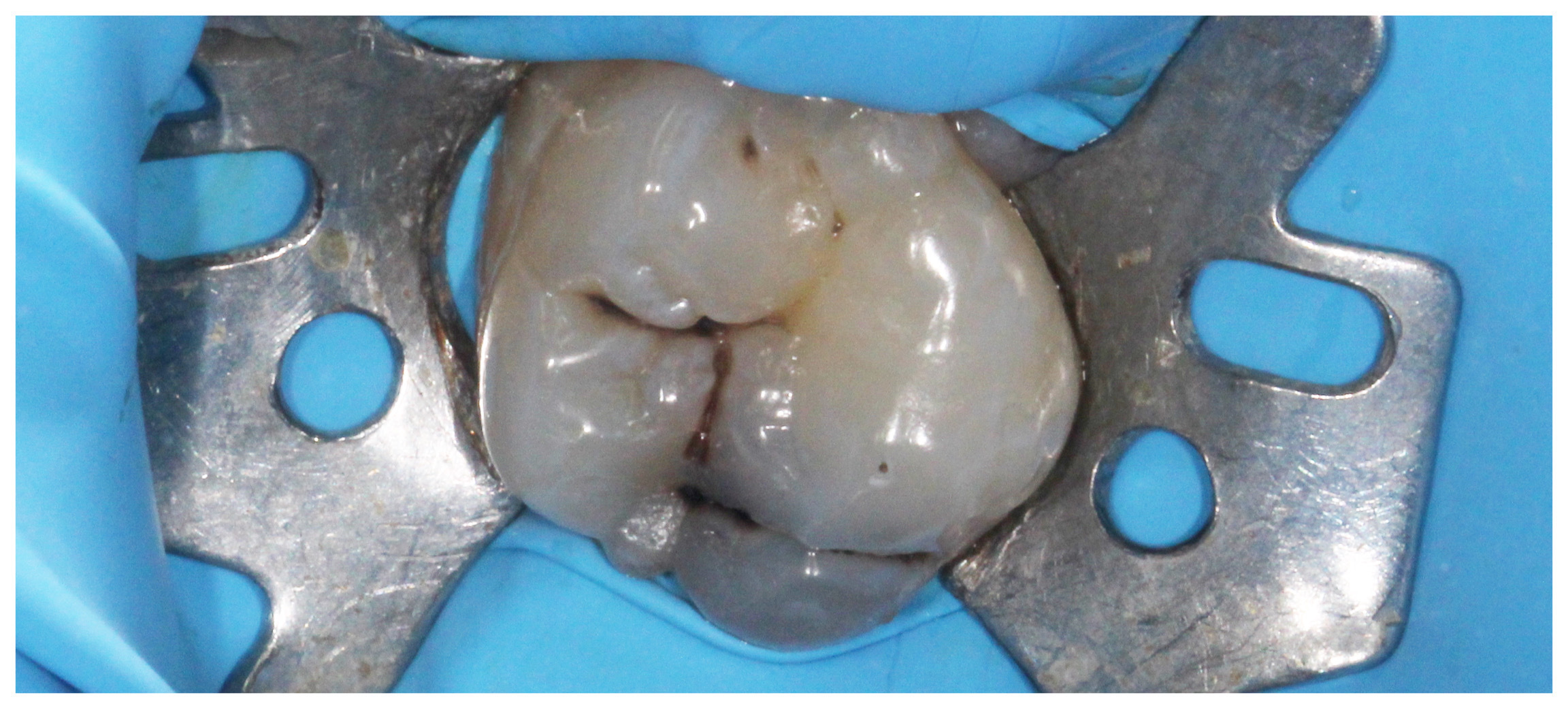
Primary caries on the upper first molar
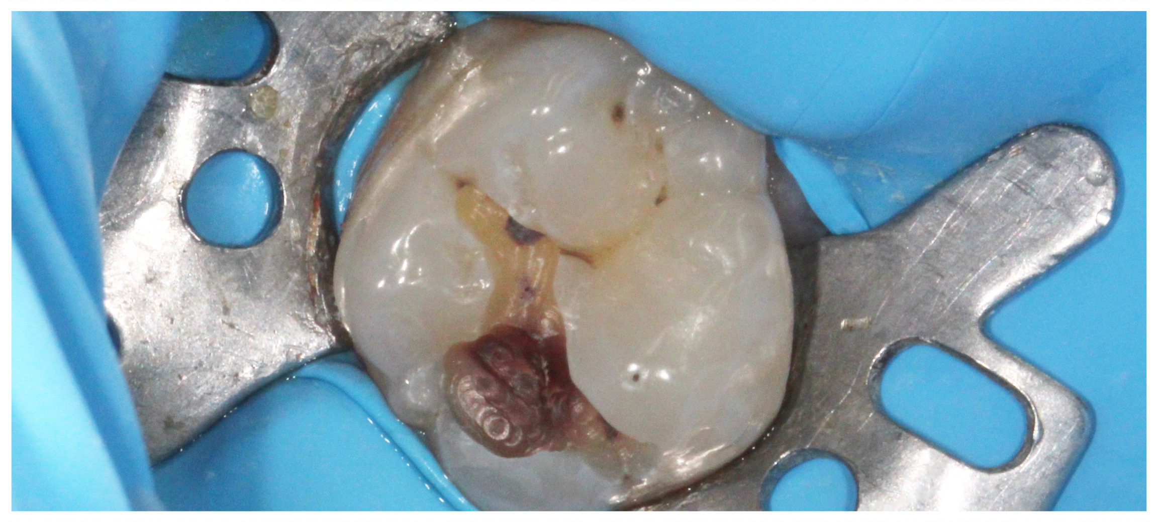
Acces to the caries
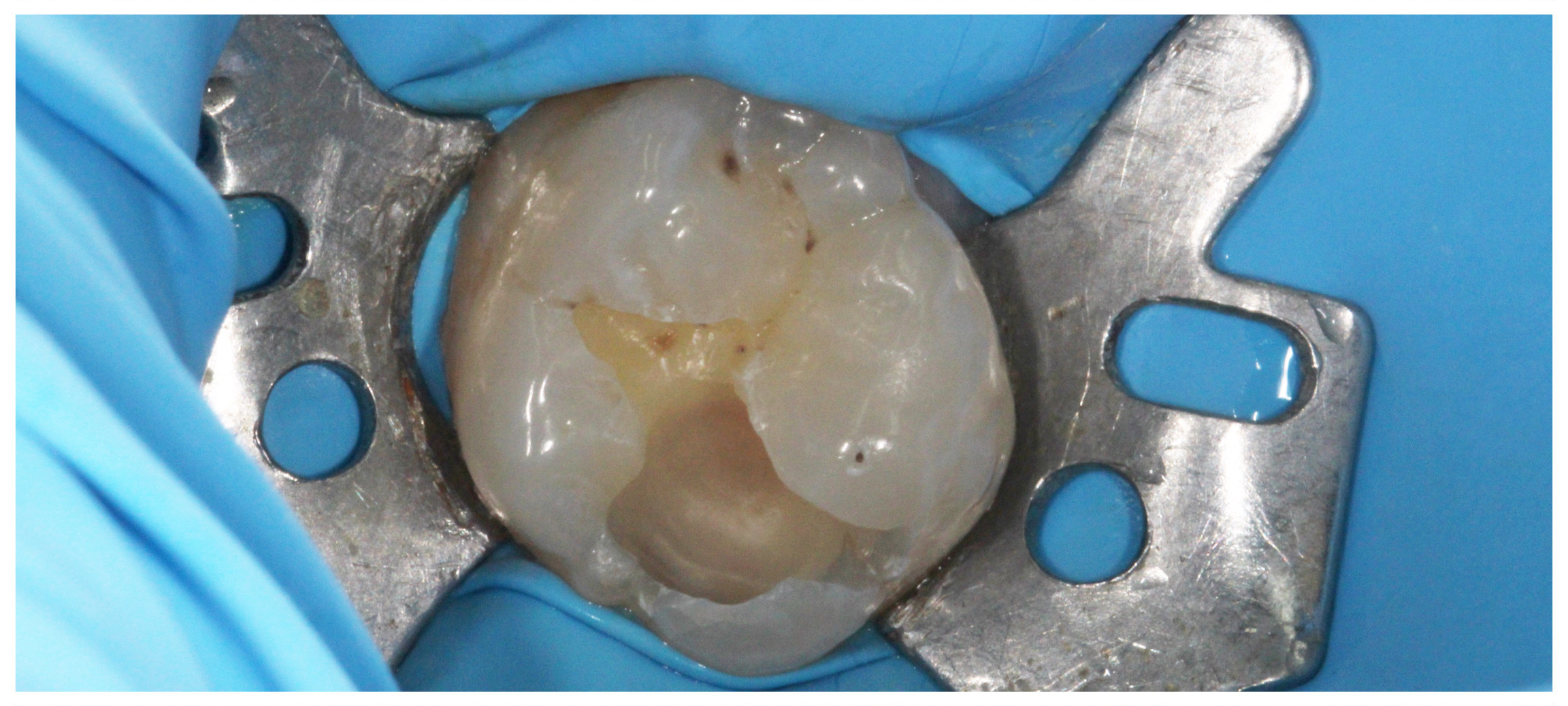
Removed caries
Photocomposite filling
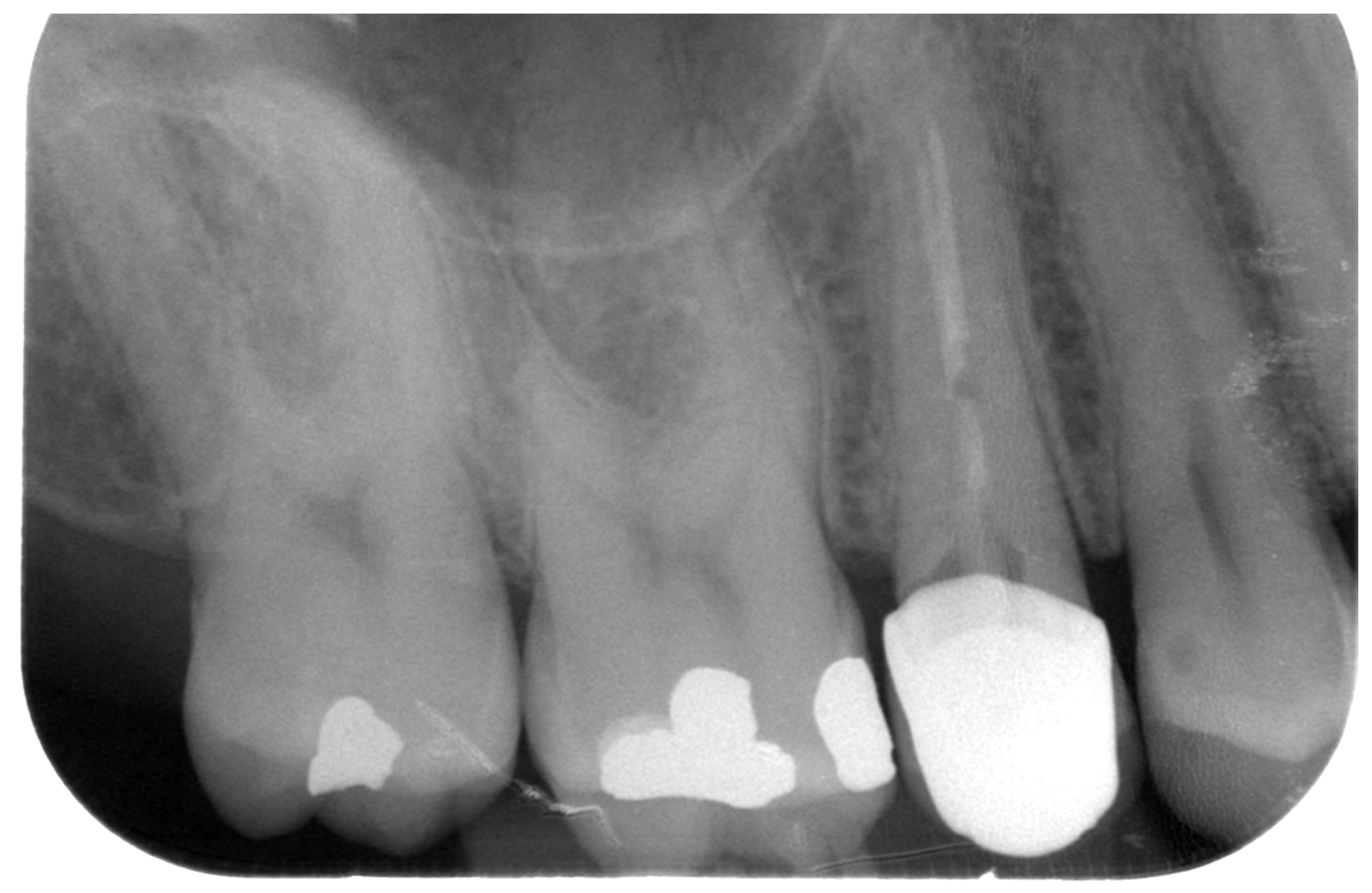
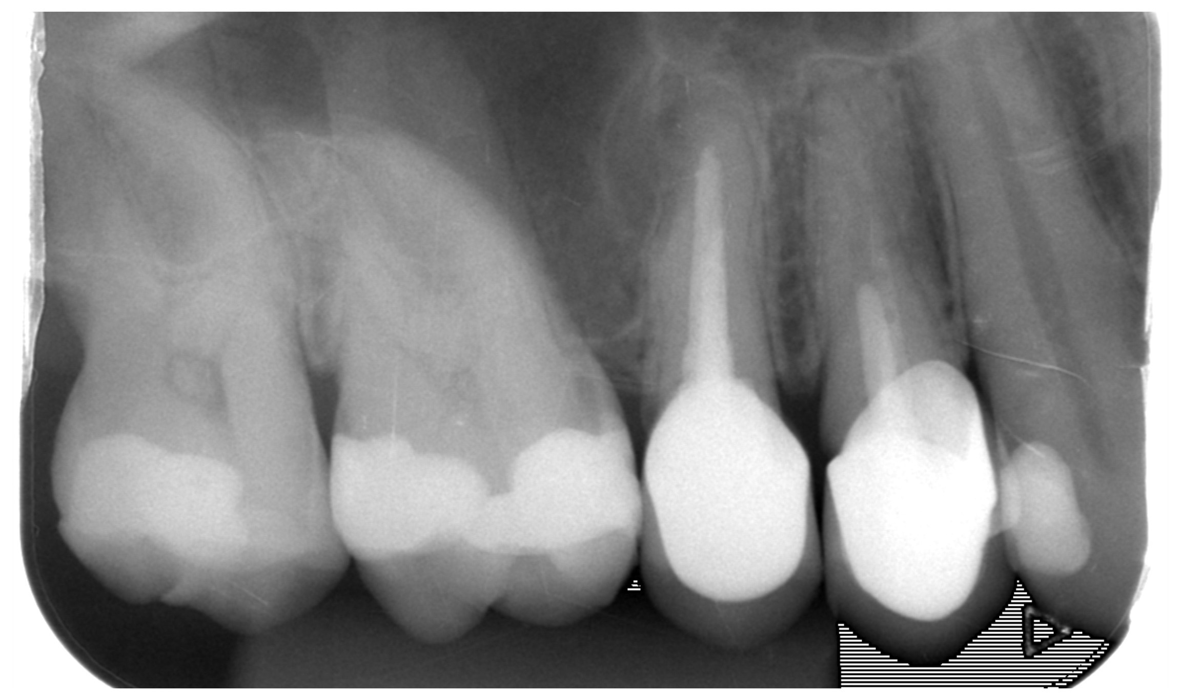
Initial X-ray on upper second premolar – inadequate root canal treatment, caries under the unexact crown, amalgam filling with caries on upper molars
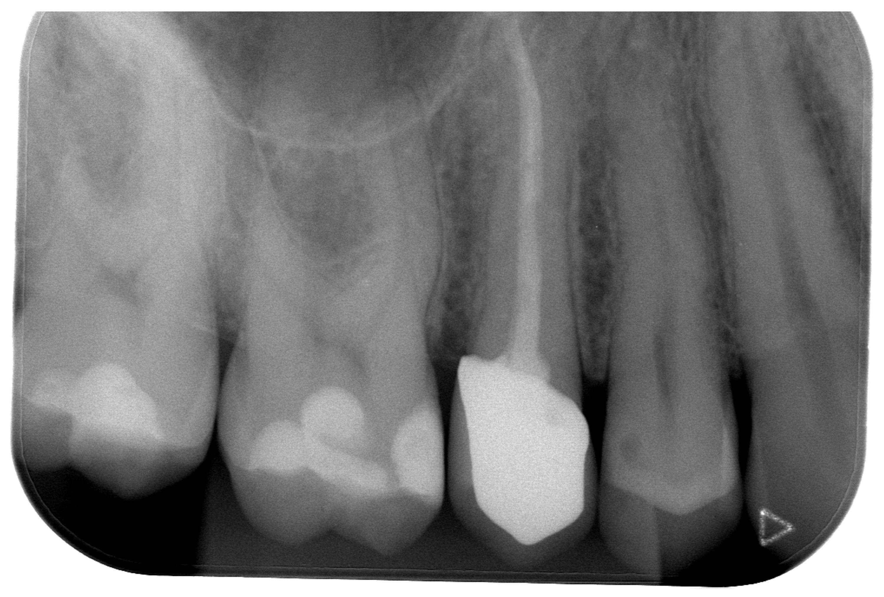
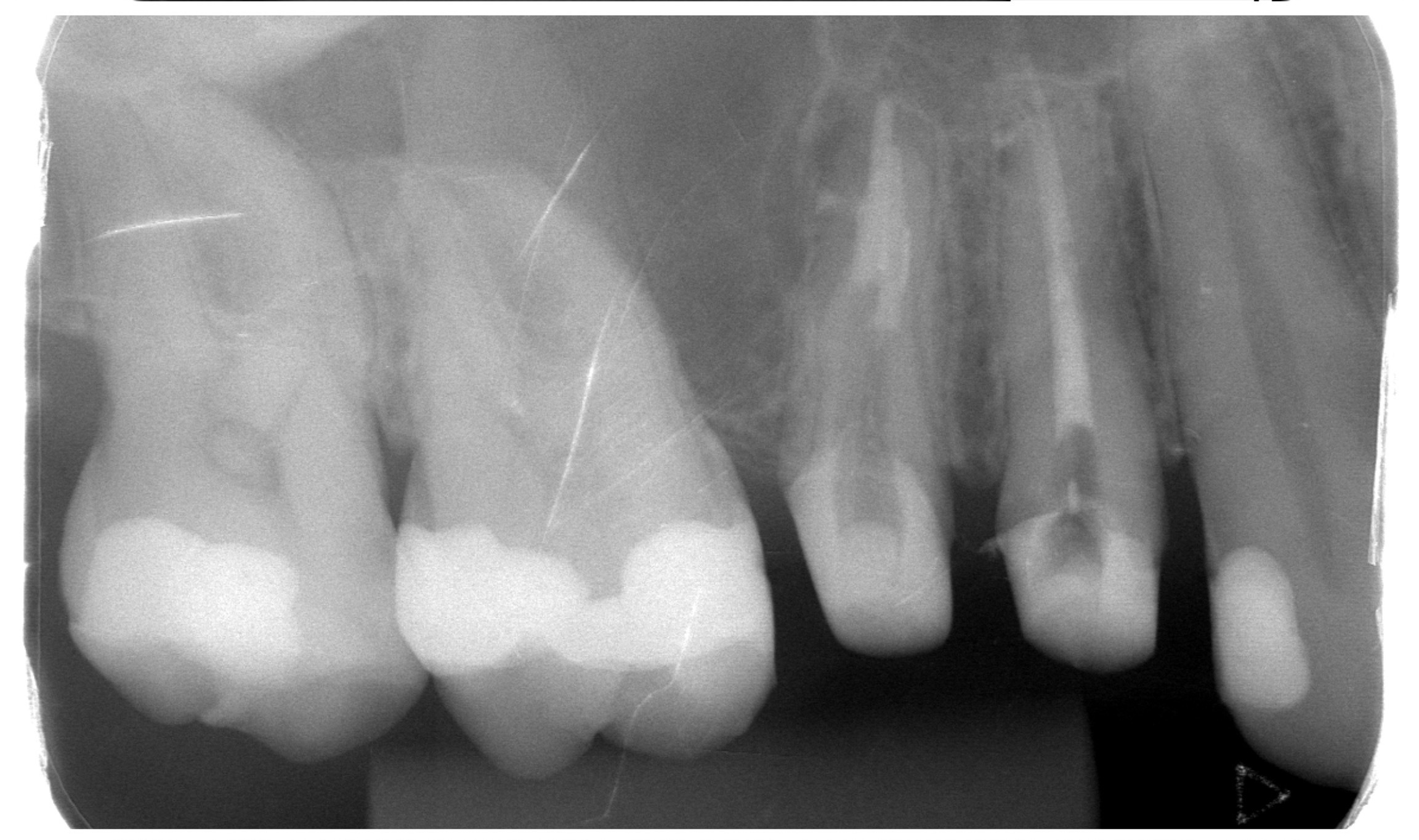
X-ray after treatment – root canals on second premolar retreated, new precise zirconium ceramic crown and new photocomposite fillings on molars
X-ray after root canal treatment of lowe premolar with periapical inflammation
Control X-ray three month later – inflammation healed (+ new photocomposite filling on molar)
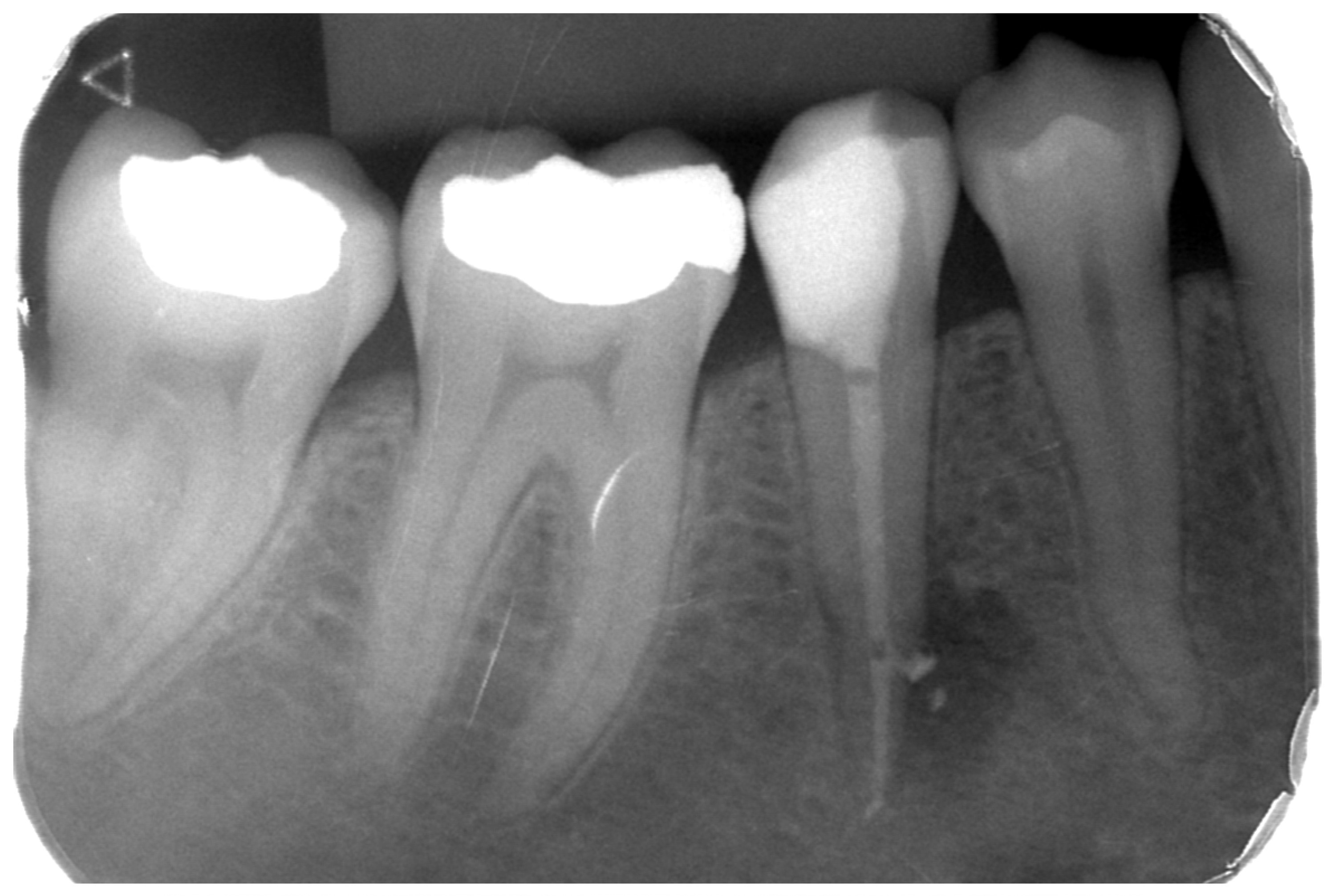
Initial X-ray – caries on lower second premolar and molars and first molar with gangrene
Control X-ray – treated root canals of lower first molar and fillings on premolar and molars